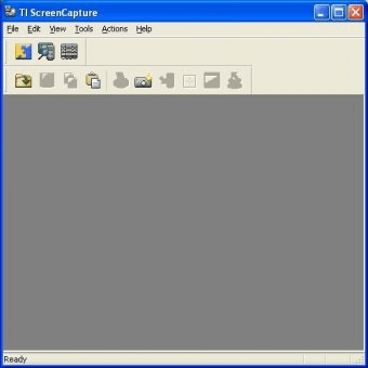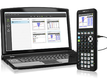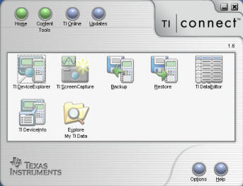

- TI CONNECT VERSION 1.6.1 INSTALL
- TI CONNECT VERSION 1.6.1 UPDATE
- TI CONNECT VERSION 1.6.1 WINDOWS 8.1
- TI CONNECT VERSION 1.6.1 WINDOWS 8
NET 4.0 is required but usually Windows 8 has this version pre-installed. For optimal performance under Windows 8 the "Activate Steinberg Power Scheme" in the Cubase/Nuendo Device Setup must be disabled for all versions older than Cubase 7 and Nuendo 6.
TI CONNECT VERSION 1.6.1 INSTALL
Please download the latest version from the website and make sure to install it prior to other Steinberg applications. Some of the compatible products require the latest eLicenser Control (eLC) version.Please make sure that you always use the latest updates available!.Older products which are not listed are not supported and consequently, no further updates are planned. For newer products, check the corresponding product pages and the system requirements on supported operating systems.
TI CONNECT VERSION 1.6.1 WINDOWS 8.1
You can find a list of Windows 8.1 compatible Steinberg applications below.
TI CONNECT VERSION 1.6.1 UPDATE
This requires an institutional login.We’d like to update you on the development and certification process regarding Windows 8.1 for current Steinberg products.

Online version available for university members only.

ProQuest Ebook Central Click for accessĪvailable from Ebook Central. ?u.ignore_date_coverage=true&rft.mms_id=9930713700301221 Ebook Central Academic Complete. Wiley ebooks (Wiley UBCM 2022-2023 Ongoing). ProQuest: Academic Complete - Click to Connect via Durham University. An e-book from Ebook Central click to view. Ebook Central Academic Complete UKI Edition. Connect to electronic book via Ebook Central. John Wiley An electronic book accessible through the World Wide Web click to view. 2.3.2 General Spore Structure2.3.3 Spore Germination 2.3.4 Polar Filament Extrusion 2.3.5 Spore Activation 2.3.6 Polar Tube Eversion and Transit from the Spore 2.3.7 Sporoplasms 2.3.8 Sporoplasm Transfer and Infection 2.3.9 Polar Tube Proteins 2.4 INTRACELLULAR MICROSPORIDIAL DEVELOPMENT AND INTERFACIAL RELATIONSHIPS 2.4.1 Type I: Microsporidia considered to have Direct Contact 2.4.2 Type II: Microsporidia with Parasite-Produced Interfacial Envelopes 2.4.3 Type III: Microsporidia with Host-Produced Interfacial Envelopes. 1.10.5 Human Pathogenic Microsporidia of the Family Tubulinosematidae1.11 STRUCTURE-RELATED TECHNIQUES IN MICROSPORIDIA RESEARCH 1.11.1 Recognizing Infected Hosts and Making Diagnostic Smears 1.11.2 Recognizing Microsporidian Spores 1.11.3 Observation and Measuring of Microsporidian Spores ACKNOWLEDGMENTS REFERENCES Chapter 2 Developmental Morphology and Life Cycles of the Microsporidia 2.1 INTRODUCTION 2.2 MICROSCOPIC IDENTIFICATION OF SPORES BY LIGHT AND ELECTRON MICROSCOPY 2.3 LIFE CYCLE 2.3.1 Phase I of the Microsporidian Life Cycle, Environmental and Infective. 1.7.4 Parasitophorous Vacuole as Envelope of Parasite Origin?1.8 CYTOLOGICAL ANOMALIES IN MICROSPORIDIA 1.9 MICROSPORIDIA-INDUCED EFFECTS ON HOST CYTOLOGY 1.9.1 Association with Host Cell Organelles 1.9.2 The Parasitophorous Vacuole as Host Cell Product? 1.9.3 Host Cell Hypertrophy 1.9.4 Infections of the Digestive Tract Epithelium 1.10 COMMENTS ON THE STRUCTURE OF SOME HUMAN OPPORTUNISTIC MICROSPORIDIA 1.10.1 Enterocytozoon bieneusi 1.10.2 Encephalitozoon Species 1.10.3 Trachipleistophora Species 1.10.4 Vittaforma corneae. 1.5 STRUCTURE OF MICROSPORIDIAN DEVELOPMENTAL STAGES1.5.1 The Spore 1.5.2 Meronts and Merozoites 1.5.3 Sporogony 1.5.4 The Sporoblast 1.5.5 The Origin of the Extrusion Apparatus 1.6 THE MATURE SPORE 1.6.1 Spore Shape 1.6.2 Spore Size 1.6.3 The Spore Wall 1.6.4 The Plasma Membrane 1.6.5 The Nucleus 1.6.6 Cytoplasmic Organelles 1.6.7 The Polar Filament 1.6.8 The Polaroplast 1.6.9 The Posterior Vacuole 1.7 ENVELOPES OF MICROSPORIDIAN ORIGIN 1.7.1 Sporophorous Vesicles 1.7.2 Exospore-Derived Envelopes 1.7.3 Sporont-Derived Sacs. Microsporidia: Pathogens of Opportunity Copyright Contents Contributors Preface Acknowledgments Chapter 1 Structure of Microsporidia 1.1 INTRODUCTION 1.2 STRUCTURAL CHARACTERS AND CLASSIFICATION OF MICROSPORIDIA 1.2.1 Microsporidia under the Light Microscope 1.3 STRUCTURAL ASPECTS OF REPRODUCTION AND LIFE CYCLES 1.3.1 Basic Life Cycle 1.3.2 Reproduction Modes 1.3.3 Sexual Processes 1.3.4 Life Cycle Types 1.4 ESSENTIALS OF MICROSPORIDIAN CYTOLOGY 1.4.1 The Plasma Membrane 1.4.2 The Nucleus 1.4.3 Mitosomes 1.4.4 Endoplasmic Reticulum 1.4.5 Ribosomes 1.4.6 Golgi Apparatus.


 0 kommentar(er)
